What Imaging Confirms the Need for Lipoma Treatment?
Lipomas are among the most common benign soft tissue growths found beneath the skin. While they are generally harmless and often painless, their evaluation becomes important when they grow in size, cause discomfort, or raise diagnostic uncertainty. In such cases, imaging plays a crucial role in confirming the nature of the lump and determining whether Lipoma Treatment in Dubai is necessary.This article explores the imaging techniques used to evaluate lipomas, how they help in diagnosis, and when they indicate the need for medical intervention.
Understanding Lipomas
A lipoma is a slow-growing, fatty lump that develops between the skin and underlying muscle. It typically feels soft, movable, and doughy to the touch. Most lipomas are small and do not require treatment unless they:
- Grow rapidly
- Become painful or tender
- Restrict movement
- Cause cosmetic concerns
- Show atypical features
In such situations, imaging is used to confirm whether the mass is a simple lipoma or something that requires further attention.
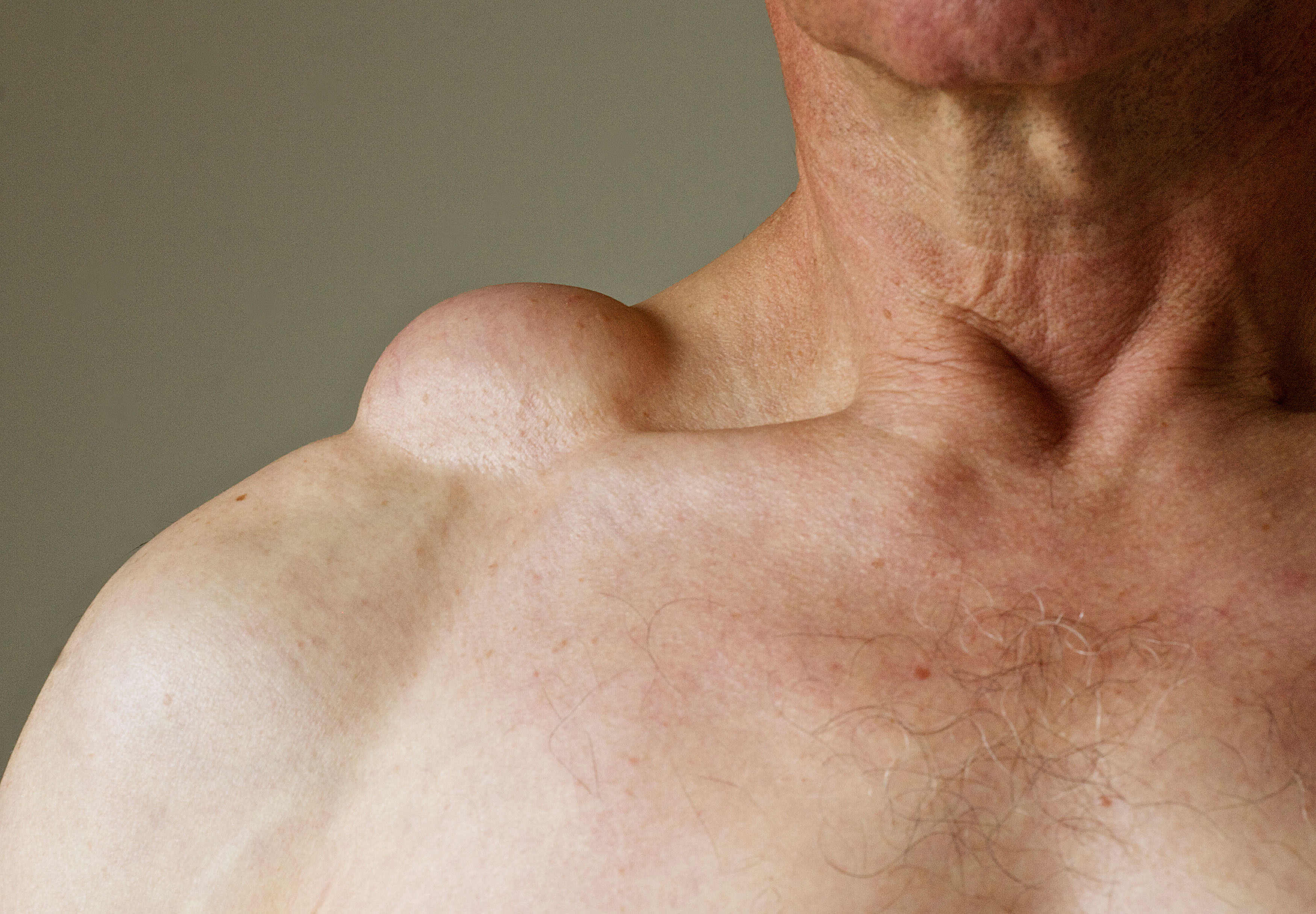
Why Imaging is Important in Lipoma Evaluation
Although many lipomas can be diagnosed through physical examination alone, imaging is essential when:
- The diagnosis is uncertain
- The lump is deep-seated or unusually large
- There are atypical symptoms
- There is suspicion of a more complex condition
Imaging helps differentiate lipomas from other soft tissue tumors, cysts, or, in rare cases, malignant growths.
Common Imaging Techniques Used
Several imaging modalities are used to evaluate lipomas. Each provides different levels of detail and is selected based on the clinical scenario.
1. Ultrasound Imaging
Ultrasound is often the first imaging tool used to assess a suspected lipoma.What it shows:
- A well-defined, homogenous, and oval-shaped mass
- Typically located within the subcutaneous tissue
- Soft and compressible structure
Why it is useful:
- Non-invasive and widely available
- No radiation exposure
- Helps confirm that the lump is fatty in nature
- Useful for distinguishing cysts from solid masses
Ultrasound is particularly helpful for superficial lipomas and is commonly used as an initial diagnostic step before considering Lipoma Treatment in Dubai.
2. Magnetic Resonance Imaging (MRI)
MRI provides a more detailed view of soft tissues and is considered one of the most accurate imaging methods for evaluating lipomas.What it reveals:
- High signal intensity on T1-weighted images
- Suppression of signal on fat-saturation sequences
- Clear borders separating the lipoma from surrounding tissues
Why it is important:
- Helps identify deep or intramuscular lipomas
- Differentiates between benign lipomas and liposarcomas
- Provides precise anatomical mapping before treatment
MRI is especially recommended when:
- The lipoma is large or deep
- There are atypical features
- Surgical planning is required
3. Computed Tomography (CT) Scan
CT scans are less commonly used than MRI but can still provide useful information in certain cases.What it shows:
- Fat-density lesions with uniform characteristics
- Clear margins in most lipomas
- Relationship with nearby structures
When it is used:
- When MRI is not available
- For evaluating deeper masses in complex anatomical areas
- In cases where other imaging results are inconclusive
CT scans help confirm the fatty composition of a mass, supporting the diagnosis of a lipoma.
Imaging Features That Suggest a Lipoma
When imaging is performed, certain characteristics strongly suggest that a lump is a benign lipoma:
- Homogeneous fat density or signal
- Well-defined, encapsulated borders
- Absence of internal septations or irregularities
- No invasion into surrounding tissues
- Slow growth pattern
If these features are present, the diagnosis of a lipoma is more likely, and treatment decisions are based on symptoms rather than urgency.
Imaging Features That May Require Further Evaluation
In some cases, imaging findings may indicate that the mass is not a simple lipoma. Red flags include:
- Thick internal septa
- Nodular or irregular components
- Heterogeneous signal intensity
- Rapid growth
- Deep tissue involvement
- Ill-defined margins
Such features may prompt further investigation to rule out other conditions. In these situations, imaging plays a vital role in guiding whether Lipoma Treatment in Dubai should proceed and what approach is appropriate.
When Imaging Leads to Treatment Decisions
Imaging not only confirms the diagnosis but also helps determine whether treatment is needed. Lipoma treatment may be recommended when:
- The lipoma causes pain or discomfort
- It interferes with daily activities or movement
- It continues to grow over time
- It is cosmetically concerning
- Imaging shows atypical or uncertain characteristics
Based on imaging findings, healthcare professionals can decide whether observation or removal is the best course of action.
Role of Imaging in Treatment Planning
If treatment is considered, imaging provides valuable guidance for:
- Determining the exact size and depth of the lipoma
- Locating its position relative to muscles, nerves, and vessels
- Planning a safe and effective removal approach
- Reducing the risk of complications during treatment
Accurate imaging ensures that the procedure is tailored to the individual case, improving outcomes and minimizing unnecessary intervention.
Conclusion
Imaging plays a critical role in confirming the diagnosis of lipomas and determining whether treatment is required. Techniques such as ultrasound, MRI, and CT scans help distinguish lipomas from other soft tissue masses, assess their characteristics, and guide clinical decisions.For individuals considering Lipoma Treatment in Dubai, imaging provides clarity, reassurance, and a structured pathway toward appropriate care. While many lipomas remain harmless and require no intervention, imaging ensures that any unusual or concerning features are properly evaluated, allowing for informed and timely decisions.